
Can an ECG Detect a Heart Attack?
 Read time: 6 mins
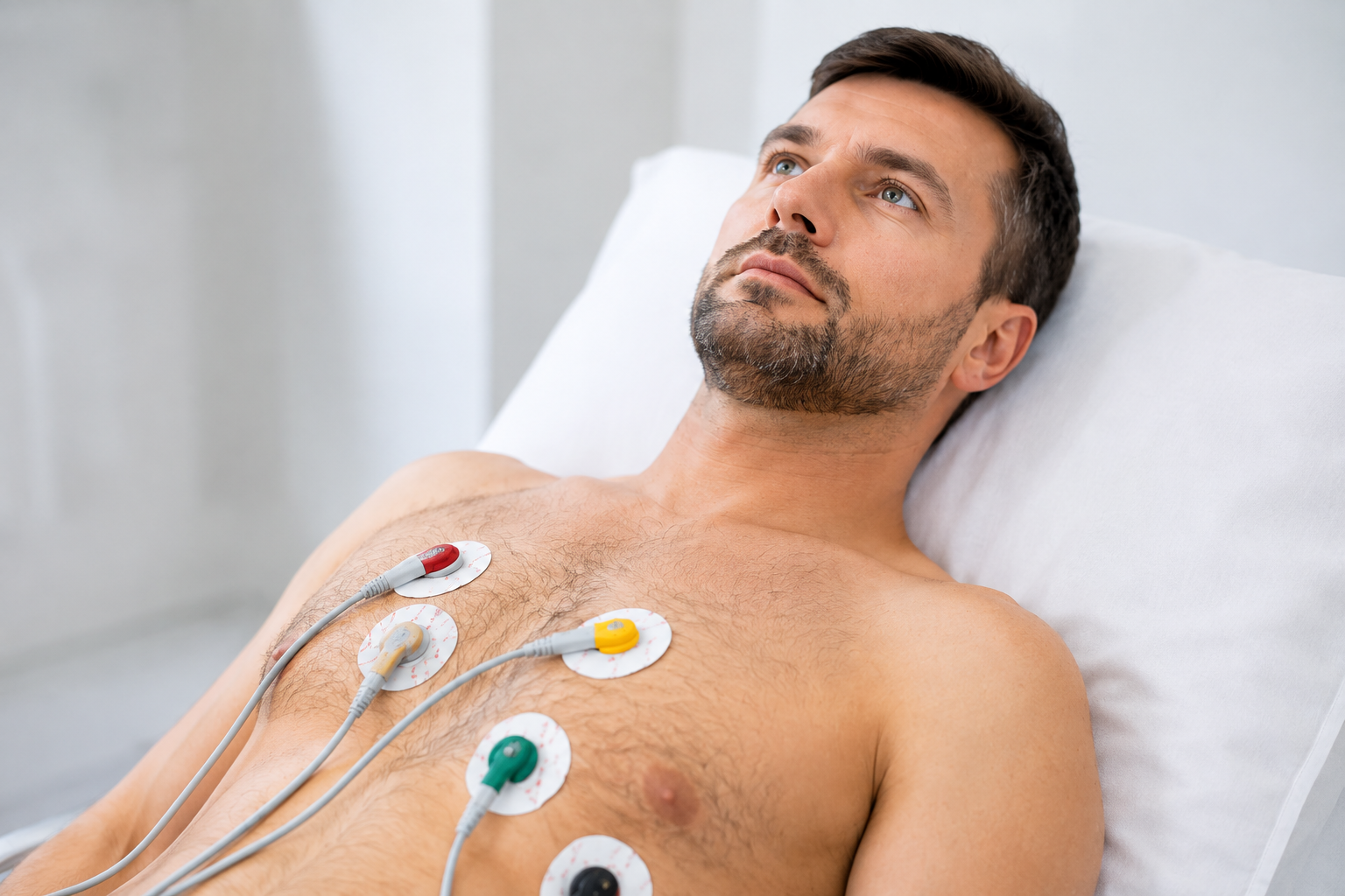
What is an ECG and how does it work
An ECG test is a simple painless test used to detect the heart’s electrical impulses. Sensors attached to the skin on the arms, legs and chest detect the electrical signals produced by your heart each time it beats. These are displayed on a screen for interpretation.
An ECG is requested if there are concerns about the heart’s structure and function, such as when people have chest pain, breathlessness, palpitations, dizziness, or loss of consciousness and as a screening test for a wide range of inherited cardiac conditions
The ECG
- Records the hearts rate and rhythm
- Helps identify abnormal heart electrical activity and irregular heart rhythm
- Highlights changes linked to reduced blood supply to the heart muscle
Importantly, an ECG measures electrical signals, not blood flow itself.
Can an ECG Detect a Heart Attack?
ECGs are used to assess the heart in both urgent and routine settings. While the test plays an important role in identifying heart problems, it isn’t always clear what an ECG can and can’t confirm when a heart attack is suspected. In this article, Dr David Brull (Consultant Cardiologist at St John & St Elizabeth Hospital) explains what is an ECG, when it can detect a heart attack, and why further tests are needed. Dr Brull is an experience cardiologist with a specialist interest in cardiopulmonary exercise testing in perioperative risks.
The Short Answer: Can an ECG Detect a Heart Attack?
Yes, an ECG can detect a heart attack, but not always.
An electrocardiogram (ECG) records the heart’s electrical activity. During a heart attacks (also known as myocardial infarction), as heart muscle is damaged, this electrical activity changes often in recognizable ways. In practice, these changes may include alterations in parts of the ECG, typically the ST segment (such as elevation or depression), that suggest the heart muscle is not receiving enough blood.
These patterns help clinicians decide whether urgent treatment is needed, but they don’t appear in every heart attack.
However, a normal ECG does not always rule out a heart attack, especially in the early stages. This is why ECGs are used alongside other tests, not on their own. All these tests can be carried out in our private cardiology clinic.
When Can an ECG Detect a Heart Attack
The diagnosis of a heart attack is based on the combination of clinical assessment, an ECG and blood tests.
A heart attack is caused by a blockage in one of the heart’s arteries (coronary arteries) This is usually due to a combination of slowly built-up fatty narrowing that develop over many years (called atheroma) in combination with a blood clot (or thrombosis). If a coronary artery blocks this leads to changes in the heart’s electrical activity often detectable on ECG, with symptoms such as chest discomfort and breathlessness and abnormalities in a series of blood test that look to detect evidence of heart muscle damage.
An ECG is particularly useful when a heart attack causes clear electrical changes, such as:
- ST segment elevation (also called ST elevation)
- Abnormal patterns in the ST segment
These changes often indicate a medical emergency requiring urgent treatment, especially when symptoms like chest pain are present. In these cases, an ECG can help doctors diagnose a heart attack quickly and start treatment immediately.
When an ECG May Not Detect a Heart Attack
A normal ECG does not always exclude a heart attack.
An ECG may appear normal when:
- The heart attack is very recent or if the affected area is small
- If there is a partial blockage of the coronary arteries, when symptoms may come and go
This is why doctors repeat ECGs and combine them with blood tests.
Why Blood Tests Are Needed To Check For Heart Attacks
If a heart attack is suspected, doctors order blood tests to check cardiac markers, usually for the troponin level. Troponin is a marker of cardiac (heart)injury), that occurs during a heart attack.
Troponin is released into the blood soon after heart muscle cells are damaged early after a heart attack. Because troponin levels rise over time, they are measured more than once, usually 1-4 hours apart. Since the diagnosis of heart attack involves a combination of clinical review, ECGs and blood tests patients may be kept in hospital for observation even if an initial result is normal, particularly when symptoms or ECG findings remain unclear or with ongoing symptoms.
If a heart attack is suspected other tests may be used
Depending on symptoms, risk, and initial results, a healthcare professional may recommend:
- Imaging tests that use sound waves (ultrasound), called echocardiography to look at heart structure and function
- X-ray based imaging to look at the heart’s blood supply, called coronary angiography ( usually as an urgent and invasive day a case procedure but this may be performed non-invasively in a CT scanner).
- Exercise ECG or stress test (often using a stationary bike or treadmill)
- 24-hour Holter monitor for continuous monitoring of heart rhythm tracking
- Chest X-ray or chest X to assess the heart and lungs
Heart Attack Risk Factors
There are many reasons why a person may suffer a heart attack. Common risk factors include:
- Increasing age
- Smoking
- High cholesterol
- High blood pressure (hypertension)
- Diabetes
- Family history of heart attack below age 60
- Sedentary lifestyle, poor diet and obesity (exacerbating many of these risk factors)
Assessment of these factors help determine the risk of future heart attacks and guide next steps.
What to Do If You Have Heart Symptoms Despite a Normal ECG
If you have prolonged episodes of chest discomfort lasting 15 minutes of more, (or shorter but recurring periods of chest discomfort), shortness of breath, dizziness, or feel something isn’t right, even with a normal ECG, seek medical help urgently. A heart attack is a medical emergency, and early assessment saves lives.
Never ignore symptoms based on one test alone.
FAQs for ECG
Who should have an ECG?
- For the investigations of chest pain, breathlessness, high blood pressure, palpitations or dizziness
- Routine pre-operative assessment, particularly before moderate or high-risk surgery
- Baseline assessment for patients starting certain medications (such as those that prolong the QT interval)
- As part of a cardiovascular health screen, especially for individuals with risk factors such as hypertension, diabetes, smoking, family history of heart disease, or elevated cholesterol
- Following a syncopal (fainting) episode
Can an ECG detect a silent heart attack?
Sometimes, but not always. An ECG may show changes that suggest a past or silent heart attack, particularly if there has been damage to the heart muscle that affects its electrical activity. However, some silent heart attacks cause minimal or no lasting electrical changes, meaning the ECG can appear normal. This is why ECG findings are reviewed alongside symptoms, medical history, and other tests such as blood work or cardiac ultrasound (echo cardiography).
How soon after symptoms should an ECG be done?
An ECG should be performed as soon as possible after heart-related symptoms begin, especially symptoms such as chest pain, shortness of breath, dizziness, or palpitations. Early ECGs can help identify urgent heart problems, but electrical changes may not appear immediately. For this reason, ECGs are often repeated over time if symptoms continue or worsen.
Is an ECG enough to rule out heart problems?
No. While an ECG is an important and widely used test, it cannot rule out heart conditions on its own. Some heart problems, including early or mild heart attacks, blood flow issues, or structural heart conditions, may not show clear changes on an ECG. Doctors use ECGs alongside blood tests, imaging, and clinical assessment to build a complete picture of heart health.
What is the role of an Exercise ECG
An exercise ECG, known as a stress test or exercise tolerance test (ETT), monitors the heart’s electrical activity during exercise (either on a treadmill or stationary bicycle).
It can be used to assess for problems with the hearts blood supply or rhythm and has a number of roles such as:
- Assessing exercise capacity and functional status — crucial for risk stratification, surgical fitness assessment, and rehabilitation planning
- Evaluating symptoms that occur only during exertion — such as exercise-induced arrhythmias, assessing for poor heart rate response (chronotropic incompetence, failure of heart rate to rise appropriately), or exercise-induced hypotension ( causing dizziness or fainting)
- Pre-participation screening — for athletes or individuals embarking on vigorous exercise programmes, particularly those with cardiovascular risk factors
The principle behind exercise testing is simple, looking for changes in the hearts blood supply ( ischaemia testing) manifest as changes on ECG that occur during stress. By increasing the heart’s workload, any underlying coronary artery narrowing limiting blood flow to the heart muscle becomes more apparent. As heart rate rises, areas of the heart supplied by narrowed arteries may become ischaemic ( lacking in blood supply) producing characteristic changes on the ECG tracing, often associated with symptoms such as chest discomfort or breathlessness.
During exercise testing the workload is gradually increased in stages according to standardized protocols using set criteria to limit the test or if a person feels unwell or unable to continue exercising. The ECG is recorded throughout exercise and into recovery period.
During the test blood pressure response, heart rate recovery, and a note of symptoms such as chest tightness or breathlessness are also recorded. An exercise test is performed under close supervision and is very safe.
Beyond detecting ischaemia, the exercise ECG provides valuable functional information. Research has consistently demonstrated that patients who can achieve higher workloads (for example, 10 METs or above) without ST-segment changes have an excellent prognosis, regardless of other risk factors
Posted on: 6 August 2026
Last updated: 6 August 2026
Tags:
Content provided by Dr David Brull - MBBS BSc MD FRCP