Key Treatments
Eye problems can be genetic or stem from an injury or trauma. Get a care plan completely tailored around your unique wants and needs, at our Eye Clinic.
If you don’t see a required treatment below, get in touch for more information.
Contact number: 020 7806 4060
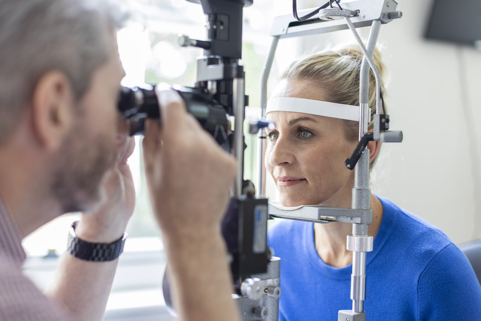
The Eye Clinic offers modern treatment techniques and cutting-edge diagnostics for all eye conditions and complaints. We have specifically designed the clinic to offer the very best care and treatment for eye problems, all under one roof. Our consultants have access to the latest diagnostic testing and fully equipped operating theatres.
At St John & St Elizabeth Hospital’s Eye Clinic, our leading consultant ophthalmologists provide expert care for a wide range of eye conditions in a calm and comfortable setting. Whether you’re experiencing vision problems or need treatment for cataracts, glaucoma, macular degeneration or other concerns, we offer fast access to consultations, diagnostics and treatment – all under one roof. Most procedures are carried out as day cases using the latest techniques, helping you return to daily life with minimal disruption.
Eye problems can be genetic or stem from an injury or trauma. Get a care plan completely tailored around your unique wants and needs, at our Eye Clinic.
If you don’t see a required treatment below, get in touch for more information.
Our Ophthalmic Consultants have a wealth of experience and expertise in diagnosing and treating eye problems.
The multidisciplinary team in the Eye Clinic consists of specialist surgeons, orthoptists, medical photographers, paediatric ophthalmologists, paediatric optometrists and nurses trained in ophthalmic procedures.
The Eye Clinic offers modern treatment techniques and cutting-edge diagnostics for all eye conditions and complaints. We have specifically designed the clinic to offer the very best care and treatment all under one roof.
The clinic has the latest technology for treating a wide range of eye conditions. Our specialists are all registered consultants within the NHS, many of them leaders in their fields.
We recognise that coming into hospital can be a stressful and an unnerving experience. At St John & St Elizabeth Hospital we are committed to the welfare of all our patients and you will find all staff will work hard to ensure your stay with us is a positive experience.
You can read articles, interviews and blogs on a range of health topics on our blog page. We frequently speak to our expert consultants and clinicians to get their specialist advice.